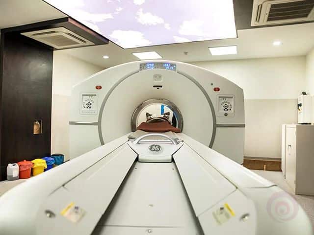
Advanced DOPA PET CT Scan for Neuroendocrine Tumors, Parkinson's Disease, Brain Disorders, Pheochromocytoma and Paraganglioma across Chandigarh, Mohali & Panchkula.
Years
Scans
Support
Reports
A F-18 DOPA PET CT Scan is an advanced Nuclear Medicine imaging test that evaluates dopamine metabolism and certain neuroendocrine tumors. Unlike routine imaging, it detects abnormal cellular activity before structural changes become obvious on CT or MRI.
A very small amount of Fluorine-18 Fluorodopa (F-18 DOPA) is injected into a vein. After a short uptake period, the PET scanner detects the radioactive tracer while the CT scan provides detailed anatomical images. The combined PET-CT images help doctors accurately localize abnormal tissue.
Useful in selected neurological disorders including Parkinsonian syndromes and dopamine pathway evaluation.
Helps identify neuroendocrine tumors, recurrent disease and metastatic lesions.
Combines metabolic PET information with detailed CT anatomy for accurate diagnosis.
Review medical history and verify the prescription.
F-18 DOPA tracer is injected through a small IV line.
Relax comfortably while the tracer circulates.
The scanner captures metabolic and anatomical images.
Nuclear Medicine specialists prepare the report for your treating doctor.
A DOPA PET CT Scan is recommended when your doctor needs detailed functional imaging for neurological disorders or specific neuroendocrine tumors. It helps detect disease earlier, evaluate recurrence, locate hidden lesions and guide treatment planning.
| Clinical Situation | Preferred Imaging |
|---|---|
| Parkinsonian Disorders | DOPA PET β |
| Pheochromocytoma | DOPA PET β |
| Paraganglioma | DOPA PET β |
| Routine Cancer Imaging | FDG PET |
| Prostate Cancer | PSMA PET |
| Neuroendocrine Tumors (Somatostatin Receptor) | DOTA PET |
Our DOPA PET CT program supports diagnosis, staging, treatment planning and follow-up for neurological and neuroendocrine disorders using advanced PET-CT technology.
Assessment of dopamine pathways in selected movement disorders and neurological conditions.
Localization of primary and recurrent neuroendocrine tumors with functional imaging.
Helps detect catecholamine-producing adrenal tumors and extra-adrenal disease.
Whole-body evaluation for suspected or recurrent paraganglioma.
Combined metabolic and anatomical imaging to identify disease spread.
Monitor treatment response and detect recurrence after surgery or therapy.
Detects metabolic changes before many structural abnormalities become obvious.
Combines PET metabolism with CT anatomy for accurate localization.
Supports surgical planning, radiation therapy and systemic treatment decisions.
Evaluates disease in multiple regions during a single examination.
Helpful when conventional imaging is inconclusive or tumor markers rise.
Non-invasive imaging with expert Nuclear Medicine interpretation.
Choosing the right imaging study depends on the suspected disease. The table below summarizes when DOPA PET CT is preferred compared with other commonly performed scans.
DOPA PET: Better for selected neuroendocrine tumors and dopamine pathway imaging.
FDG PET: Preferred for many common cancers with high glucose metabolism.
DOPA PET: Useful in selected NETs and catecholamine-producing tumors.
DOTA PET: Ideal for somatostatin receptor-positive neuroendocrine tumors.
DOPA PET: Not intended for prostate cancer imaging.
PSMA PET: Gold-standard PET imaging for prostate cancer evaluation.
DOPA PET: Functional imaging showing metabolic activity.
MRI: Excellent anatomical detail for brain and soft tissues.
DOPA PET: Detects abnormal cellular function.
CT: Demonstrates structural abnormalities and anatomy.
PET identifies abnormal metabolism while CT precisely localizes the abnormality, improving diagnostic confidence.
| Feature | DOPA PET | FDG PET | DOTA PET | MRI | CT |
|---|---|---|---|---|---|
| Functional Imaging | β | β | β | β | β |
| Structural Imaging | With CT | With CT | With CT | β | β |
| Brain Disorders | Excellent | Limited | Limited | Excellent | Moderate |
| Neuroendocrine Tumors | Excellent | Variable | Excellent | Limited | Limited |
| Routine Cancer Imaging | Selected Cases | Excellent | Selected Cases | Limited | Moderate |
Following the preparation instructions provided by your treating physician and imaging center helps achieve the best image quality. Individual instructions may vary depending on your condition and medications.
Verify your appointment, identity and clinical history.
F-18 DOPA is administered through a small intravenous line.
Relax comfortably while the tracer distributes within the body.
The scanner acquires metabolic PET images along with CT images.
Your images are interpreted by a Nuclear Medicine specialist.
Always inform the imaging team if you are pregnant, breastfeeding, have recently undergone another nuclear medicine procedure, or have specific medical conditions that may affect preparation or interpretation.
We combine advanced PET-CT technology, experienced Nuclear Medicine specialists and patient-focused care to provide accurate diagnostic imaging for patients across Chandigarh, Mohali and Panchkula.
High-quality imaging with modern PET-CT equipment for precise diagnosis.
Reports interpreted by experienced Nuclear Medicine physicians.
Priority scheduling with same-day appointment support when available.
Convenient online report access and image sharing.
Guidance for eligible insurance and reimbursement documentation.
Parking, wheelchair accessibility and convenient city location.
Conveniently located in Sector 35B, Chandigarh, serving patients from the entire Tricity and nearby regions.
MRI Chandigarh
SCO 315β316, First Floor
Sector 35-B, Chandigarh β 160035
Call 8699572364 or WhatsApp us to schedule your appointment and receive preparation guidance.
Book NowDesigned to reassure patients while highlighting modern PET-CT imaging and quality-focused care.
Years of Diagnostic Experience
PETβCT Studies
Appointment Assistance
Digital Reporting
"The staff explained every step clearly and the entire DOPA PET scan process was smooth and comfortable."
"Appointment booking was quick and the report helped my doctor plan further treatment confidently."
"Professional team, modern equipment and excellent support from registration to reporting."
Common questions from patients in Chandigarh, Mohali and Panchkula.
A DOPA PET Scan is a specialized PET-CT study using F-18 Fluorodopa to evaluate dopamine metabolism and selected neuroendocrine tumors.
It helps diagnose, stage and monitor selected neurological conditions and neuroendocrine tumors.
FDG PET evaluates glucose metabolism, while DOPA PET targets different biological pathways useful in specific diseases.
Apart from a small IV injection, the procedure is generally painless.
Most appointments take 2β3 hours including preparation, tracer uptake and imaging.
Preparation varies. Follow the instructions provided by your treating physician and imaging centre.
In many cases hydration is encouraged unless instructed otherwise.
It is generally considered safe when performed under appropriate medical supervision.
The scan uses a controlled amount of radioactive tracer and CT radiation appropriate for the examination.
Pregnancy should always be discussed with your doctor before scheduling the scan.
Inform the imaging team so they can provide individualized guidance.
It is used for selected movement disorders and neuroendocrine tumors including pheochromocytoma and paraganglioma.
It may help evaluate dopaminergic pathways in selected clinical situations.
Yes, it can be valuable in selected neuroendocrine tumors depending on clinical indication.
Bring previous reports, prescriptions and follow the preparation instructions given before your appointment.
Yes, previous CT, MRI or PET studies help comparison.
Yes, images are interpreted by a Nuclear Medicine specialist and a report is issued.
Availability depends on the clinical situation and reporting workflow.
Qualified Nuclear Medicine physicians review the images.
It may be performed when clinically indicated under specialist supervision.
Comfortable clothing is generally recommended.
Most patients can resume routine activities unless advised otherwise.
Appointments are available for patients from Chandigarh, Mohali, Panchkula and nearby areas.
Pricing varies by indication and centre. Contact the imaging centre for the latest quotation.
Experienced teams, modern equipment and quality reporting help support accurate diagnosis.
Yes, it may help identify recurrent disease when clinically appropriate.
Yes, DOPA PET CT is typically performed on the advice of a treating specialist.
Tell the imaging team about all current medicines and supplements.
Yes, but preparation may differ. Follow the instructions given by your doctor.
Inform the staff beforehand so they can discuss ways to improve comfort.
Some PET-CT studies may include CT contrast depending on the indication.
No. DOPA PET and MRI provide complementary information.
No. Tissue diagnosis may still be required in many situations.
Accuracy depends on the disease being investigated and should be interpreted with clinical findings.
Reporting times vary depending on the case complexity.
Most patients can travel after completing the examination unless advised otherwise.
Follow the instructions provided by the Nuclear Medicine team.
No. The tracer decays naturally and is eliminated from the body.
Most patients can resume their normal diet unless instructed otherwise.
Patients with appropriate neurological or neuroendocrine indications.
Yes, appointments are available for patients across the Tricity.
Coverage depends on your insurer and policy.
Prescription, previous reports and valid identification.
A family member can usually accompany you to the centre.
Discuss the findings with your treating physician.
Please confirm parking availability while booking.
Many centres provide WhatsApp booking assistance.
Call 8699572364 to schedule an appointment.
Contact the centre to reschedule.
Experienced team, advanced imaging and patient-focused service.
Call 8699572364 to discuss your prescription, preparation instructions and appointment availability.
Book AppointmentDOPA PET CT is a highly specialized Nuclear Medicine imaging technique that helps physicians detect abnormal dopamine metabolism and evaluate selected neuroendocrine tumors. It provides valuable metabolic information that complements MRI and CT findings for more accurate diagnosis and treatment planning.
Evaluates dopaminergic pathways in selected patients with suspected Parkinsonian disorders.
Useful in differentiating certain movement disorders when clinically indicated.
Detects primary and recurrent neuroendocrine tumors with high diagnostic accuracy in selected cases.
Helps localize adrenal tumors producing excess catecholamines.
Whole-body imaging for suspected or recurrent paragangliomas.
Can assist in evaluating selected patients with recurrent or metastatic disease.
Advanced metabolic imaging for selected pediatric neuroendocrine tumors.
Supports evaluation of selected brain tumors and helps differentiate active disease from treatment-related changes in appropriate clinical settings.
Useful when conventional CT or MRI findings are inconclusive and recurrence is suspected.
May help identify the source of disease in carefully selected patients.
Provides additional metabolic information when blood tumor markers remain elevated.
Helps evaluate response after surgery, chemotherapy, targeted therapy or radiation treatment.